
|
by Lauren Lin Induced pluripotent stem cells (or iPSC) are stem cells that are made by reprogramming already specialized adult cells into a pluripotent state that is similar to that of an embryonic stem cell. As a result, these induced pluripotent stem cells are able to differentiate into any cell type in the body. This technology was developed by Shinya Yamanaka and Kazutoshi Takahashi at Kyoto University in 2006 when they infected adult skin cells from mice with viruses to introduce 24 genes that they believed to be necessary for cells to behave like embryonic stem cells. Later on, they were able to identify that there were only four genes, each of them encoding for a transcription factor, that were needed to reprogram adult cells into pluripotent cells. These genes were Oct4, Sox2, Myc, and Klf4.
iPS cells are now widely used within many areas of research, but the iPS cell lines cultured in different labs don’t seem to be consistent, as many papers have reported results that other researchers haven’t been able to replicate. Additionally, there is still research being done to investigate which types of adult cells are able to be reprogrammed, which cells the iPSC can differentiate into, and whether or not reprogramming can take place without Myc, since it has the potential to turn cells cancerous. It has also been discovered that iPSCs aren’t exactly the same as embryonic stem cells, since they hold onto a certain kind of “epigenetic memory.” This means that the cells have retained the chemical changes associated with whichever adult cell they originated from. However, some scientists don’t believe that this epigenetic memory will end up interfering with the results of research conducted with iPSC. When iPSC techniques were first developed, researchers originally thought that it would be primarily used for regenerative medicine. It was thought that they could reprogram a person’s skin or blood cells into iPSCs, then differentiate these cells into whichever cell type was needed to treat a disease. For example, people with neurological disorders could have new neurons made to treat their condition. The idea that iPSCs could be used for regenerative medicine was especially exciting because it seemed to be able to avoid both the problem of immune rejection and the ethical issues that arise from using stem cells from embryos in therapy. In 2013, Masayo Takahashi worked with Yamanaka to develop stem cell treatments for retinal diseases. Takahashi and her team reprogrammed skin cells from patients with an eye condition called age-related macular degeneration (AMD) into iPSCs, which were then used to make retinal pigment epithelium (RPE) cells. Sheets of RPE cells were implanted into a patient in 2014, and the progression of the patient’s macular degeneration seemed to stop. However, before a second trial was started, they found a few genetic changes in the second patient’s iPS cells and RPE cells. It wasn’t conclusive whether or not the mutations were cancerous, but the trial was halted and only resumed in June 2016. When Takahashi’s work was stopped, other researchers developing iPS-cell-based therapies also put their projects on hold. Now that the clinical trials for AMD are set to begin again, researchers hope to start new clinical trials that examine the use of iPSCs for other diseases, such as Parkinson’s disease. Currently, iPS cells are heavily used in research, especially for newly developed drugs and the progression of human diseases. The ability to culture iPS cells and to differentiate these cells into any body cell has allowed researchers to culture human tissues that would have previously been difficult to access. Additionally, while clinical trials have experienced setbacks, iPSC technology has been continuously refined and used for other studies. For example, some researchers have been using CRISPR-Cas9, a gene-editing tool, to introduce mutations into iPS cells to look at the effects of the mutation and to compare the mutated cell lines with control cell lines. The research that studied the link between the Zika virus and microcephaly, a condition in which an infant is born with a head that is abnormally small, used the ability of iPSCs to model early human development. Dr. Guo-li Ming used cortical neural progenitor cells, iPSCs, and immature neurons to discover that the cells that go on to form the brain’s cortex are “potentially susceptible to the virus, and their growth could be disrupted by the virus.” Although it will still take a very long time to understand certain diseases and to develop new drugs or cell therapies, iPSCs are valuable tools that researchers can use, either as models for human tissue or as treatments themselves. iPSCs have already contributed to many scientific advances, and there may be potential uses for them that have yet to be explored.
0 Comments
by Hannah Latour 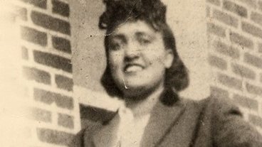 Being able to culture and grow cells is integral to many areas of biomedical research. This technique has allowed scientists to develop vaccines, map genes and even clone organisms. Regardless of the use, every cell line must be sourced from somewhere. Rebecca Skloot’s novel “The Immortal Life of Henrietta Lacks” delves into the origin of HeLa – the oldest and most utilized cell line in science. However, this story is about more than cells. Racism, patient consent, tissue ownership and research ethics all intersect at the forefront of HeLa.
Henrietta Lacks was born in 1920 in Roanoke, Virginia. She had five children, and shortly after the birth of her youngest child, was diagnosed with an aggressive form of cervical cancer. Being a poor, black tobacco farmer in the 1950s didn’t leave Henrietta with a lot of treatment options. Hospitals in the United States were still racially segregated, and the medical services offered to black patients were severely lacking. The public wards of Johns Hopkins hospital in Baltimore, Maryland became one of the few places black patients could seek medical attention. This is where Henrietta sought treatment, and where HeLa was born. After receiving a diagnosis of cervical cancer, Henrietta began radiation treatment. At this time, Dr. George Gey, a doctor and cell biologist was trying to grow the world’s first immortal cell line in his lab at Johns Hopkins. While undergoing radiation, a biopsy was taken (without permission) from Henrietta’s cervical tumor – and given to the Gey lab for study. While Henrietta died from her disease in 1951 – her cancer cells (taken from her tumor) were growing rapidly. A lab technician labelled a vial containing the growing cells “HeLa” – short for Henrietta Lacks, and this was how the cell line got its name. There are several theories about why Henrietta’s cells thrived, while so many others failed to be cultured. HeLa cells are immortal, meaning they can divide an infinite number of times. Most cells can only divide a limited number of times before they die, so it has been suggested that the cancerous nature of the cells imparted this property. HeLa cells survived so well in culture, that they were sent to other research labs – being transported through postal mail and remaining alive at the end of the journey. Eventually, a HeLa production centre was established – supplying HeLa cells to researchers worldwide. While this “factory” started off as a not-for-profit, it was taken over by Microbiological Associates which began producing the cells for profit. While all of this was going on, Henrietta’s family was unaware of how her cells had revolutionized scientific research, let alone the fact they had been commercialized. It wasn’t until 1975 that Henrietta’s family discovered her cells were a multimillion-dollar industry. A few years prior to this, researchers from Johns Hopkins approached Henrietta’s children to obtain blood samples, that would be used to conduct further research regarding HeLa. Informed consent was not obtained for this, as the children were never given the full story about HeLa or its use in research. Additionally, portions of Henrietta’s medical records were also published without her family’s permission. The contribution of HeLa to the scientific community has been massive. HeLa was used to develop the polio vaccine, grow viruses and test the effects of radiation and other toxins. HeLa has increased our knowledge of cancer and cellular processes in general. It seems ironic that despite this impact on medicine, Henrietta’s children cannot afford health insurance. The majority of Skloot’s novel chronicles Deborah Lacks, Henrietta’s youngest daughter. Deborah is plagued by a laundry-list of medical conditions, many related to the chronic stress of living in border-line poverty. Deborah is also haunted by the fact that she remembers almost nothing of her mother, yet her cells live on in laboratories around the world. The issue of patient consent and tissue collection span beyond HeLa. It is estimated that over 307 million tissue samples from over 178 million people are currently stored in tissue banks, and that the majority of these samples were taken without the knowledge of the people they came from. There are different perspectives on whether an individual still “owns” their tissue after it has been removed from their body. In 1951, it wasn’t illegal for Henrietta’s cells to be used without her permission. Today, bio-specimens in tissue banks can be used for research without patient consent, provided that the specimens cannot be traced back to a specific individual. However, the legislation regarding the ownership of human tissue continues to be debated. Skloot’s novel highlights the complexities of ethics in research, in the context of racial discrimination. Would Henrietta’s family have been kept in the dark if they were privileged and white? Maybe, but maybe not. Ultimately, Skloot’s narrative puts a human face on a scientific issue, illustrating how interlinked the two entities are. Image courtesy of: http://www.sydsvenskan.se/images/Beg-1m_Hs42QsuhWqSO5EyYxjy4.jpg |
Categories |
 RSS Feed